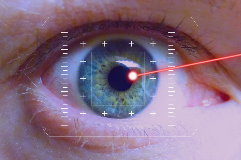
United States Military Tests Wavefront-Guided PRK

With more than 30,000 to 40,000 members of the United States military choosing laser vision correction procedures to improve their vision each year, there is an emphasis on clinical research to examine the efficacy and safety of various procedures available.

A somewhat novel laser vision correction technique, called wavefront-guided PRK (Photorefractive Keratectomy), was recently studied by the United States Navy.

Wavefront-guided PRK implements the newest computer-generated mapping technology to generate a digital image of the patient’s eye. Once the eye is mapped out, the laser applied to the cornea’s surface is guided by the three-dimensional model to improve the eye’s focus based on the unique visual characteristics of the patient’s eye. Even the tiniest structure issues affecting eyesight are revealed through Wavefront-Guided PRK.

The development of wavefront vision mapping allows visual disorders to be measured, which otherwise could not be identified in the past. Laser diagnostic systems are setting a new standard by providing an exact level of measurement and correction. This feat was impossible with earlier systems.

Led by the U.S. Naval Medical Center, 167 patients enrolled had vision prescriptions of up to -8D of nearsightedness and up to -4D of astigmatism. The patients underwent wavefront-guided PRK. Research findings showed:

  • Patient satisfaction was very high, with 98.8 percent reporting entirely or very satisfied with their vision
  • 99.4 percent of patients tested at 20/20 or better vision six months following the procedures
  • 91.9 percent of patients tested 20/16 vision six months after the procedure
  • Fewer than four percent of patients reported night vision symptoms six months post-op
  • Less than one percent experienced adverse reactions

This novel research shows a massive body of evidence supporting laser vision correction procedures proving safety and effectiveness. 

Rohr Eye & Laser Center offers the most advanced technology available to provide personalized and extraordinary care to our patients. Whether your goal is to maintain or improve your natural vision, we are here to help you. Call us at 877-579-0202 or visit https://www.michiganlasik.com/ to schedule an appointment today.

Written by the digital marketing staff at Creative Programs & Systems: www.cpsmi.com.

Retinal Transplants Ready for Human Trial

A major cause of hereditary human blindness, Retinitis Pigmentosa (RP), has been resolved in rat trials by researchers at the RIKEN Center for Biosystems Dynamics Research (BDR) in Japan. Degenerated rat retinas received transplants and removal of grafted cells, which resulted in improved connections to host retinas and better light-responsiveness.

Study leader Michiko Mandai said, “We have previously reported work using mouse embryonic- and induced pluripotent stem cell – (iPES)-derived retinas in a mouse model of retinal degeneration.”

However, the deputy project leader in the Laboratory for Retinal Regeneration at RIKEN BDR said, “As far as we know, this is the first report of the timed removal of particular grafted cells being shown to improve retinal function in an animal model using tissue from human stem cells.”

Since the retinal sheets were created from human stem cells, the novel feat can now be tested in human clinical trials, according to the study authors in the January 21, 2022 edition of iScience.

“Since retinal ganglion cells form optic nerves to send the signal to the brain, a higher light responsiveness could theoretically lead to improved sensitivity to light or better resolution in visual function. Genetic modification in human stem cell-derived retinas showed a substantial functional improvement [in light responsiveness], compared with wild-type graft retinas. Depending on the retinal network of the transplanted area, light responsiveness of retinal ganglion cells may be correlated with increased signaling to the brain, so could lead to an increased light sensitivity or better resolution. Additionally, we were able to observe detailed host-graft synapse formation in the absence of graft bipolar cells, which previously was difficult. A frequently asked question in retinal cell therapies concerns whether retinal ganglion cell responses are really originated from the graft photoreceptors. The presence of multiple synaptic markers at the host-graft cell contact site suggests formation of some synapses between these cells and indicates that graft photoreceptor light responses may well be transmitted to host retinal cells. We can now move forward to applying this strategy in clinical studies, which we expect to improve clinical outcomes and be generally useful in stem cell-based therapies targeting retinal degeneration. If the clinical grade ISL1-deleted stem cell-derived retinas can first pass extensive safety testing, including for tumorgenicity, we estimate that this may take 4-5 years. While we are currently targeting end-stage disease eyes with stem cell based therapy, we think this strategy could also be applied to those with remaining central vision, to help increase the pericentral visual field or delay disease progression,” Mandai said.

Rohr Eye & Laser Center offers the most advanced technology available to provide personalized and extraordinary care to our patients. Whether your goal is to maintain or improve your natural vision, we are here to help you. Call us at 877-579-0202 or visit https://www.michiganlasik.com/ to schedule an appointment today.

Written by the digital marketing staff at Creative Programs & Systems: www.cpsmi.com.

New Nerve Cell Discovered in Retina

A new type of neuron, or nerve cell, has been discovered in the retina by scientists at the John A. Moran Eye Center at the University of Utah.

A complex group of neurons communicate with each other in the central nervous system, relaying sensory and motor information. These interneurons are an essential aspect of communication. This finding marks a breakthrough for scientists who are working toward a better understanding of the central nervous system. By identifying and classifying all neurons, researchers can more easily distinguish between different aspects.

A research team led by Ning Tian, Ph.D., identified the interneuron in the mammalian retina. Tian said, “Based on its morphology, physiology, and genetic properties, this cell doesn’t fit into the five classes of retinal neurons first identified more than 100 years ago. We propose they might belong to a new retinal neuron class by themselves.”

The new neuron is dubbed Campana after a handbell, resembling its shape. Campana cells are able to transmit visual signals in the retina, but the exact function is yet to be determined.

Tian said, “In the brain, persistent firing cells are believed to be involved in memory and learning. Since Campana cells have a similar behavior, we theorize they could play a role in prompting a temporal ‘memory’ of a recent situation.”

Rohr Eye & Laser Center offers the most advanced technology available to provide personalized and extraordinary care to our patients. Whether your goal is to maintain or improve your natural vision, we are here to help you. Call us at 877-579-0202 or visit https://www.michiganlasik.com/ to schedule an appointment today.

Written by the digital marketing staff at Creative Programs & Systems: www.cpsmi.com.

Blind Woman Sees Shapes Thanks to Scientists

A team of researchers from the University Miguel Hernandez (Spain), the Netherlands Institute of Neuroscience (Netherlands), and the John A. Moran Eye Center at the University of Utah (USA) recently published details about how they were able to successfully restore vision in a woman using a brain implant.

The scientists oversaw a series of tests with the blind volunteer to eventually recreate her vision. First, a neurosurgeon implanted a microelectrode array into her visual cortex, which was composed of 100 microneedles intended to stimulate neurons near the electrodes. The woman wore eyeglasses outfitted with a mini video camera which translated the data collected and sent to the brains electrodes. Finally, the brain simulated the image by surrounding neurons to produce white areas of light known as “phosphenes.”

A former science teacher, the woman had been blind for 16 years. No complications were seen post-op, and scientists concluded the implant did not affect brain function. 

Lines, shapes, and letters were visible to the blind woman. The researchers developed a game to help her strengthen and recognize the different phosphene shapes. Because of her extensive involvement and unique insight, the woman became a co-author of the study.

Professor Eduardo Fernandez said, “These results are very exciting because they demonstrate both safety and efficacy and could help to achieve a long-held dream of many scientists, which is the transfer information from the outside world directly to the visual cortex of blind individuals, thereby restoring a rudimentary form of sight. Although these preliminary results are very encouraging, we should be aware that there are still a number of important unanswered questions and that many problems have to be solved before a cortical visual prosthesis can be considered a viable clinical therapy.”

Professor R. A. Normann, a study co-author, said, “It could allow them to identify a person, doorways, or cars. It could increase independence and safety. That’s what we’re working toward.”

Professor P. Roelfsema, a co-author in the study, said, “This new study provides proof-of-principle and demonstrates that our previous findings in monkey experiments can be translated to humans. This work is likely to become a milestone for the development of new technologies that could transform the treatment of blindness.”

Rohr Eye & Laser Center offers the most advanced technology available to provide personalized and extraordinary care to our patients. Whether your goal is to maintain or improve your natural vision, we are here to help you. Call us at 877-579-0202 or visit https://www.michiganlasik.com/ to schedule an appointment today.

Written by the digital marketing staff at Creative Programs & Systems: www.cpsmi.com.

Research Proves LASIK Adverse Events are Rare

Ophthalmology is a scientific field that is continuously performing research and studies based on the human eye. LASIK eye surgery is an imperative aspect of ophthalmology that presents patients with more benefits than risks.

A recent study published in the American Journal of Ophthalmology outlined researchers’ data analysis from roughly 37,000 laser vision correction patients who underwent either LASIK (31,921) or PRK (5,016) between July 2014 and June 2016. Data analyzed included eye examinations prior to the surgery, procedure reports, and follow-up exams.

Data on adverse events were also recorded, such as those that negatively impacted patients’ visual outcomes. Out of 37,000 patients, 34 experienced adverse side effects; 70 percent were characterized as severe. With less than one percent of patients experiencing a significant unfavorable result, each procedure showed:

  • LASIK (31,921 patients): 0.4 percent of patients experienced unfavorable results
  • PRK (5,016 patients): 0.7 percent of patients experienced unfavorable results

Of those with serious adverse effects, roughly 80 percent still achieved 20/20 (or higher) vision without glasses or contacts.

With over 20 million procedures performed throughout the United States over 20 years, research papers number greater than 7,000. Experts study vision correction effects in-depth to ensure they are safe and effective for patients. Based on the large number of patients country-wide, LASIK and PRK are excellent, viable options to improve vision for suitable candidates. 

To improve your vision through LASIK, contact the ophthalmology experts at Rohr Eye & Laser Center today.

Rohr Eye & Laser Center offers the most advanced technology available to provide personalized and extraordinary care to our patients. Whether your goal is to maintain or improve your natural vision, we are here to help you. Call us at 877-579-0202 or visit https://www.michiganlasik.com/ to schedule an appointment today.

Written by the digital marketing staff at Creative Programs & Systems: www.cpsmi.com.

What’s the Best Age to Get LASIK?

How old do you have to be to get LASIK? The short answer: Laser vision correction procedures such as LASIK are approved for people 18-years-old and above. For an in-depth examination of the reasoning behind this, keep reading.

First, we need to examine ocular maturity and what that means regarding development. The eyes are one of the last organs to fully mature; in some people, it can take up to age 20, but for most, they are fully grown by 13. This feat, known as “achieving ocular maturity,” is considered when a patient’s eyeglass prescription remains stable for a year or more. With a consistent prescription, the likelihood of nearsightedness or farsightedness progressing is low.

Second, your vision correction options can be gauged based on how your experience with glasses or contacts has been. Most eye care professionals want to ensure you make the right decision and prefer to personally analyze you based on your habits, compromises, and vision goals. Annual eye exams are imperative to understanding whether LASIK is right for you.

Finally, it’s essential to understand that LASIK permanently reshapes the cornea, the surface of your eye. For this reason, LASIK procedures are done on people over the age of 18. The treatment has a better chance of success throughout the years if the eyes are fully developed before the procedure.

The aging process never ceases, however. While LASIK is effective for younger candidates, when people reach the age of 40, a condition called presbyopia occurs. This is categorized by a loss of flexibility in the natural lens inside your eye and can be corrected with another procedure or by simply wearing reading glasses.

While LASIK lasts for years – decades, even – it’s essential to keep up with annual visits and connect with your doctor when necessary. If your eyes aren’t mature, it’s a good idea to have a thorough evaluation to determine when you are a good candidate. If you are over 18 and have a stable prescription, your eye care professional will help you ascertain whether you are a good candidate for LASIK.

To learn more about how LASIK can improve your life by easing the stress of wearing contacts or glasses, contact the eye care professionals at Rohr Eye & Laser Center today.

Rohr Eye & Laser Center offers the most advanced technology available to provide personalized and extraordinary care to our patients. Whether your goal is to maintain or improve your natural vision, we are here to help you. Call us at 877-579-0202 or visit https://www.michiganlasik.com/ to schedule an appointment today.

Written by the digital marketing staff at Creative Programs & Systems: www.cpsmi.com.

Maintaining Senior Eye Health

Aging affects every part of your body, even your eyes. Age-related farsightedness, or presbyopia, occurs when the lens inside your eye begins to harden. Books and other close objects become challenging to read or see clearly. Some other age-related disorders include:

  • Age-related Macular Degeneration
  • Cataracts
  • Glaucoma
  • Diabetic Retinopathy
  • Dry Eye
  • Floaters
  • Changes to Peripheral Vision

Roughly half of all age-related visual loss can be managed through early diagnosis and treatment. Ensure you get routine eye exams to help prevent some of the complications listed above. Additionally, there are plenty of things you can do at home to protect your eye sight. 

Diet: Well-balanced eating habits help your body in more ways than one. Your eyes are significantly affected by the foods you choose to eat. The following nutritious foods can help avert vision loss:

  • Vitamin A: Carrots, spinach, kale, egg yolks, dairy
  • Vitamin C: Citrus fruits, juices, broccoli, potatoes, green peppers
  • Vitamin E: Whole grains, eggs, sunflower seeds, avocado
  • Fatty Acids: Coldwater fish (mackerel, rainbow trout, salmon, etc.), corn oil, sunflower oil
  • Lutein: Kale, spinach, broccoli, brussels sprouts, corn
  • Zinc: Poultry, red meat, fish, dairy, whole grains

Quit smoking: The sooner, the better. Besides causing a plethora of cardiovascular and lung-related diseases, smoking greatly increases the chances of developing age-related macular degeneration, cataracts, and diabetic retinopathy.

Exercise: All you need is at least 20 minutes a day to stimulate your entire body by increasing blood flow. The optic nerve and retina are included in this circulatory invigoration.

Sunglasses: Ultraviolet rays emitted from the sun can cause cataracts, retina damage, and skin cancer near your eyes. Always wear sunglasses when outside.

To schedule an eye exam or learn about LASIK for seniors, contact the professionals at Rohr Eye & Laser Center today.

Rohr Eye & Laser Center offers the most advanced technology available to provide personalized and extraordinary care to our patients. Whether your goal is to maintain or improve your natural vision, we are here to help you. Call us at 877-579-0202 or visit https://www.michiganlasik.com/ to schedule an appointment today.

Written by the digital marketing staff at Creative Programs & Systems: www.cpsmi.com.

The Eyes Are a Window into the Soul – and Alzheimer’s Disease

A new study shows that the eyes are telltale signs for understanding diseases of the brain. Scientists at the UCSF Weill Institute for Neurosciences found that retinal scans can distinguish vital changes in blood vessels that might provide an early sign of Alzheimer’s disease. These scans provide insight into how one of the most common Alzheimer’s risk genes (APOE4) gives way to the disease.

Fanny Elahi, MD, Ph.D., assistant professor of neurology, member of the UCSF Memory and Aging Center (MAC) and lead author of the study, said, “The most prevalent genetic risk for Alzheimer’s disease is a variant of the APOE gene, known as APOE4. We still don’t fully understand how this variant increases risk of brain degeneration, we just know that it does, and that this risk is modified by sex, race, and lifestyle. Our research provides new insights into how APOE4 impacts blood vessels and may provide a path forward for early detection of neurodegenerative disease.”

The effects of APOE4 capillaries in the brain have been studied in mice. Elahi has long suspected these tiny blood vessels might play a crucial role in Alzheimer’s disease due to their important functions through the blood-brain barrier, such as delivering nutrients and oxygen, ridding the body of waste, and monitoring immune system responses. Elahi says damage to these blood vessels could lead to multiple issues, including the protein buildup and cognitive decline seen in Alzheimer’s patients. Since we cannot  visualize individual capillaries in living peoples’ brains, Elahi focused on the eyes instead.

Through a non-invasive eye scan, APOE4-associated capillary changes were detected in humans. Light-penetrating tissue shares biology with the brain, so researchers believe the retina might help establish APOE4 variants and how similar capillaries inside the brain are affected.

After analyzing the retinal scans, a reduced capillary density in APOE4 carriers was found, signifying an effect that increased with age. The team also compared the abnormalities found in the retinal scans to brain perfusion and found that people with higher density retinal capillary density also had greater brain blood flow.

Elahi said, “This is the first time that we have demonstrated in living, asymptomatic humans that the smallest blood vessels are affected in APOE4 gene carriers. That’s important because it suggests that the increased risk of brain degeneration and Alzheimer’s disease in APOE4 carriers might be through its effect on blood vessels. This is just the beginning. But the implications for early detection and possible intervention can be significant in combatting Alzheimer’s disease and other neurodegenerative disorders. It’s much harder to regenerate neurons than to stop their degeneration from happening in the first place. Similar to cancer, early detection can save lives.”

Rohr Eye & Laser Center offers the most advanced technology available to provide personalized and extraordinary care to our patients. Whether your goal is to maintain or improve your natural vision, we are here to help you. Call us at 877-579-0202 or visit https://www.michiganlasik.com/ to schedule an appointment today.

Written by the digital marketing staff at Creative Programs & Systems: www.cpsmi.com.

Eight Underlying Causes of Night Blindness

Night blindness, or the inability to see at night, is generally a symptom of some serious vision issues. Also known as nyctalopia, it’s important to get your eyes checked regularly to ensure everything is functioning properly. If you notice you can’t see as well in dim light while driving or adjusting from outdoors to indoors, you might have night blindness.

Symptoms of night blindness include reduced contrast sensitivity, difficulty seeing people, places, or things outdoors at night or in a dim-lit area, trouble driving at night, excessive squinting at night, or difficulty adjusting from darkness to light.

Here are some underlying factors that can cause night blindness:

  1. Vitamin A: this vital vitamin helps keep your cornea (thin layer in front of your eye) clear. It’s also an essential component of rhodopsin, a protein that lets you see in low-light conditions. Though uncommon in North America, deficiency of Vitamin A can induce night-blindness.
  2. Glaucoma: the actual eye disease itself, along with the medications used to treat it, can cause night blindness. Glaucoma is associated with pressure build-up in the eye that damages the optic nerve.
  3. Cataracts: a buildup of protein clouds the eye’s lens, which leads to impaired vision at night or in poor lighting conditions.
  4. Diabetic retinopathy: damage to the eyes’ blood vessels and nerves can result in vision loss, including challenges with nighttime sight.
  5. Myopia: also known as nearsightedness, makes distant objects appear blurry; patients describe a starburst effect around night lights.
  6. Keratoconus: this irregularly shaped cornea can result in blurry vision and sensitivity to light which can worsen at night.
  7. Usher Syndrome: a genetic condition that causes both hearing and vision loss, night blindness is also an effect.
  8. Retinitis Pigmentosa (RP): this progressive genetic eye disease can be associated with other conditions and lead to night blindness and peripheral vision loss.

After a proper diagnosis, treatments for night blindness can be mitigated by your eye care professional. There is no proven way to prevent night blindness.

If you are experiencing night blindness, we can help. Contact us and schedule an appointment today.

Rohr Eye & Laser Center offers the most advanced technology available to provide personalized and extraordinary care to our patients. Whether your goal is to maintain or improve your natural vision, we are here to help you. Call us at 877-579-0202 or visit https://www.michiganlasik.com/ to schedule an appointment today.

Written by the digital marketing staff at Creative Programs & Systems: www.cpsmi.com.

Eye Color Genetics are More Complicated than Previously Thought

According to a new study published in Science Advances, an international team of researchers led by King’s College London and Erasmus University Medical Center Rotterdam proved there are 50 new genes for eye color. In the most extensive genetic study of its kind to date, the researchers genetically analyzed almost 195,000 people throughout Europe and Asia.

Eye diseases such as pigmentary glaucoma and ocular albinism, where eye pigment levels play a role, will be better understood with the new research. Additionally, researchers discovered eye color in Asians with different shades of brown is genetically similar to eye color in Europeans ranging from dark brown to light blue.

Previous research identified a dozen genes linked to eye color, and scientists thought that eye color variations were controlled by a single gene or two. Typically, the perception was that brown eyes were more common/dominant than blue eyes.

Dr. Pirro Hyski, a co-senior author at King’s College London, said, “The findings are exciting because they bring us a step closer to understanding the genes that cause one of the most striking features of human faces, which has mystified generations throughout our history. This will improve our understanding of many diseases that we know are associated with specific pigmentation levels.”

Dr. Manfred Kayser, a co-senior author at Erasmus University Medical Center Rotterdam, said, “This study delivers the genetic knowledge needed to improve eye color prediction from DNA as already applied in anthropological and forensic studies, but with limited accuracy for the non-brown and non-blue eye colors.”

For all of your eye care needs, contact the experts at Rohr Eye & Laser Center today.

Rohr Eye & Laser Center offers the most advanced technology available to provide personalized and extraordinary care to our patients. Whether your goal is to maintain or improve your natural vision, we are here to help you. Call us at 877-579-0202 or visit https://www.michiganlasik.com/ to schedule an appointment today.

Written by the digital marketing staff at Creative Programs & Systems: www.cpsmi.com.